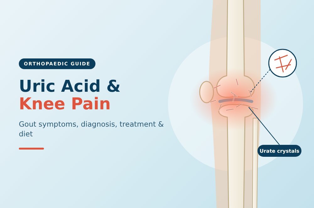
Yes, uric acid can cause knee pain, and it does so in a very specific way. When uric acid levels in the blood stay elevated for too long, needle-shaped crystals form and settle inside joints. The knee is one of the more commonly affected joints, and when those crystals trigger inflammation, the pain can be sudden, severe, and difficult to mistake for anything else.
This article explains how high uric acid and knee pain are connected, what gout in knee symptoms feel like, how it is diagnosed, what treatment options are available, and which foods to avoid for high uric acid to reduce flare frequency.
How Uric Acid Causes Knee Pain
Uric acid is a natural waste product formed when the body breaks down purines, compounds found in certain foods and produced by the body itself. Normally, the kidneys filter and excrete it through urine. When kidneys cannot keep up, or when the body produces too much, uric acid accumulates in the bloodstream. This state is called hyperuricaemia.
When uric acid levels exceed the saturation point, monosodium urate crystals begin to form. These crystals tend to settle in cooler, peripheral joints, and the knee is a frequent site. Once inside the joint space, the crystals provoke an intense inflammatory response. White blood cells rush in and attempt to engulf the crystals, releasing inflammatory chemicals that cause rapid swelling, heat, and severe pain.
According to the NICE guideline NG219 (2022), gout is the most common inflammatory arthritis, caused by deposition of monosodium urate crystals in joints and soft tissues due to chronic hyperuricaemia.
One important and often misunderstood point: not everyone with high uric acid and knee pain has gout. Some people have elevated serum urate without ever developing crystal deposition. Conversely, a normal blood uric acid level during a flare does not rule gout out, because uric acid is temporarily sequestered as crystals inside the joint during an attack.
Gout in Knee Symptoms
Gout in knee symptoms are usually distinctive enough to raise a strong clinical suspicion, even before any tests are done.
A typical flare tends to come on quickly, often overnight or in the early hours of the morning. Someone who went to bed with a mildly uncomfortable knee may wake up to find it severely painful, visibly swollen, warm, and red. Moving the joint at all can be excruciating.
The main uric acid knee pain symptoms during a flare include:
- Sudden, severe pain in the knee joint, often described as throbbing or crushing
- Visible swelling around the knee
- Warmth and redness of the overlying skin
- The joint feeling tender even to a light touch or the weight of bed linen
- Stiffness that makes bending or straightening the knee difficult
A gout flare in the knee typically resolves within one to two weeks, even without treatment, though it is very uncomfortable while it lasts. Without intervention to lower uric acid levels over the long term, further flares become increasingly likely, and the intervals between them tend to shorten.
Knee pain due to uric acid can sometimes be confused with septic arthritis, which is a joint infection. Both cause a hot, swollen, very painful knee. Septic arthritis is typically accompanied by fever and systemic unwellness. Gout can also be confused with pseudogout, which involves calcium pyrophosphate crystals rather than urate crystals and most commonly affects the knee. Distinguishing between these requires clinical assessment and, sometimes, joint fluid analysis.
When Gout Becomes a Longer-Term Knee Problem
Acute gout flares are painful but episodic. The concern with untreated or undertreated high uric acid and knee pain is what happens over years rather than days.
Chronic tophaceous gout develops when urate crystals accumulate as deposits called tophi in and around joints, tendons, and soft tissues. Tophi can appear as firm lumps around the knee, elbow, or fingers. Inside the knee, persistent crystal deposits cause ongoing low-grade inflammation and, over time, progressive joint damage. The cartilage erodes, and the joint can develop the kind of structural changes seen in advanced arthritis.
Patients who repeatedly experience knee pain due to uric acid without adequate treatment may eventually develop chronic gouty arthritis of the knee. This can look similar on imaging to osteoarthritis or rheumatoid arthritis, and distinguishing it accurately requires a proper clinical history and targeted investigation.
At this stage, the relationship between gout and knee osteoarthritis becomes clinically relevant. Untreated crystal deposition accelerates cartilage breakdown. For patients already managing moderate knee arthritis, recurrent gout flares can significantly hasten the progression of joint damage.
Diagnosing High Uric Acid and Knee Pain
When a patient presents with a hot, swollen knee, diagnosing the specific cause is the essential first step. Uric acid knee pain symptoms can overlap with other conditions, so investigations matter.
Serum Uric Acid Test
A blood test measuring serum uric acid is the standard starting point. A normal range is typically considered below 360 micromoles per litre (or around 6 mg/dL). NICE guidance notes that the level may appear normal during an acute flare, as uric acid shifts into the joint as crystals, so a normal result during a flare does not definitively exclude gout.
Joint Fluid Aspiration
Drawing fluid from the swollen knee with a needle (arthrocentesis) and examining it under a polarised light microscope is the gold standard for diagnosis. Needle-shaped monosodium urate crystals are visually distinct from other crystal types and from infection. This procedure also simultaneously relieves some of the pressure in the joint, which can reduce pain.
Imaging
X-rays of the knee may show joint erosion in chronic gout. Ultrasound can detect crystal deposits and a double-contour sign on cartilage surfaces. Dual-energy CT scanning is particularly sensitive for identifying urate crystal deposits, though it is not required in straightforward cases. These investigations become more relevant when the diagnosis is uncertain or when assessing the extent of joint damage.
For patients managing knee arthritis alongside gout, your orthopaedic surgeon may recommend clinical examination of the knee and imaging to assess the degree of structural joint involvement alongside the crystal arthritis.
Uric Acid Knee Pain Treatment
Uric acid knee pain treatment has two distinct goals: relieving an acute flare, and reducing uric acid over the long term to prevent future attacks and joint damage.
Managing an Acute Flare
NICE guidance (NG219, 2022) recommends three first-line options for treating an acute gout flare: an NSAID (nonsteroidal anti-inflammatory drug) taken with a proton pump inhibitor to protect the stomach, colchicine, or a short course of oral corticosteroids.
Colchicine works by interfering with the inflammatory response to urate crystals and is most effective when taken early in a flare. NSAIDs reduce joint inflammation and pain. Corticosteroids are useful when NSAIDs or colchicine are not tolerated or contraindicated. The right choice depends on the individual’s other medical conditions and medications. Always consult a doctor before starting any treatment.
Resting the affected knee, applying ice wrapped in a cloth, and elevating the leg can also help manage pain during a flare alongside prescribed medicines.
Long-Term Urate-Lowering Therapy
This is where most gout management falls short. NICE notes that only around one third of people with gout actually receive urate-lowering therapy (ULT), despite it being the treatment that changes the course of the disease.
Allopurinol is the standard first-line ULT. It reduces the production of uric acid by the body. Febuxostat is an alternative for those who cannot tolerate allopurinol. The target is to bring serum urate below 360 micromoles per litre. ULT is typically a lifelong treatment. When started, it can paradoxically trigger a flare as existing crystals begin dissolving and mobilising, so colchicine is often prescribed alongside it during the initial adjustment period.
For patients whose uric acid knee pain has progressed to structural joint damage requiring surgical consideration, gout control must be optimised before any planned knee replacement surgery or knee arthroscopy to reduce surgical and post-operative risk.
Foods to Avoid for High Uric Acid
Diet plays a supporting role in managing high uric acid and knee pain. The foods to avoid for high uric acid are largely those high in purines, which the body breaks down into uric acid. Dietary changes alone rarely control gout in patients with significantly elevated uric acid, but they do reduce flare frequency and support medical treatment.
Foods to Limit or Avoid
Organ meats such as liver, kidney, and sweetbreads have the highest purine content and should be avoided entirely.
Red meat (beef, lamb, pork) contributes significantly to uric acid production. Portion size matters. Limiting to occasional, moderate servings is generally advised rather than complete elimination.
High-purine seafood including sardines, anchovies, mussels, scallops, and mackerel should be restricted. Not all fish is equally problematic, and some varieties are lower in purines.
Alcohol, particularly beer, is a significant trigger. Beer contains both purines and compounds that impair uric acid excretion. Spirits also raise uric acid levels. The UK Gout Society notes that a purine-rich diet can trigger a five-fold increase in gout attacks.
Sugary drinks and high-fructose corn syrup are worth particular attention. Fructose is rapidly metabolised into purines by the body, sharply increasing uric acid levels. Fizzy drinks, fruit juices with added sugar, and many processed foods contain high-fructose corn syrup and should be reduced.
Processed and ultra-processed foods such as packaged snacks and cured meats often contain hidden purines and other compounds that worsen metabolic health.
What to Eat Instead
- Low-fat dairy products may actually lower uric acid levels
- Vegetables, including most high-purine vegetables such as spinach and mushrooms, do not appear to increase gout risk in the way animal-based purines do
- Cherries and cherry extract have shown some evidence of reducing flare frequency in small studies
- Staying well-hydrated helps the kidneys excrete uric acid more efficiently
- Whole grains, legumes (in moderation), and plant-based proteins are generally suitable
Diet modifies rather than cures gout. Patients who continue to have flares despite dietary changes should discuss urate-lowering medication with their doctor or rheumatologist.
Research Insight: Dr Amyn Rajani on Knee Pain and Joint Inflammation
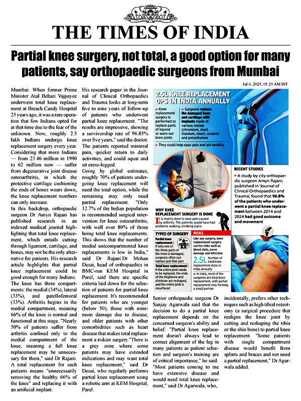
Dr Amyn Rajani is a Consultant Knee, Shoulder, and Hip Surgeon with over 25 years of experience. He practices at Breach Candy Hospital, Saifee Hospital, and PD Hinduja Hospital in Mumbai, and is a reviewer for the Journal of Arthroplasty and the Indian Journal of Orthopaedics.
One of his published case studies is directly relevant to the complexities of knee pain diagnosis. His 2021 paper “The Black Knee: A Case Report”, published in the Journal of Orthopaedic Case Reports, documented an unusual presentation of knee joint pathology with distinctive joint changes visible at arthroscopy. The case highlighted how knee pain and joint appearances can sometimes reflect underlying systemic or metabolic conditions that are not immediately obvious, reinforcing the importance of thorough investigation before assuming a cause.
This is relevant for patients with uric acid knee pain symptoms because gout is one of several metabolic joint conditions that can produce atypical or confusing presentations at surgery. Identifying the underlying cause accurately, whether it is crystal deposition, mechanical wear, infection, or ligament damage, directly shapes the treatment strategy.
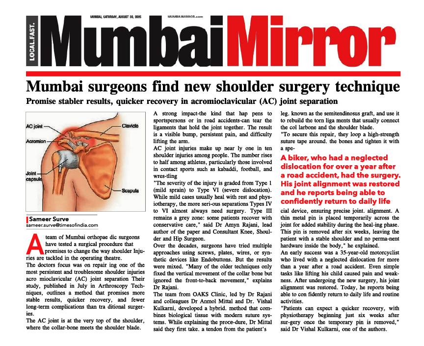
Dr Rajani’s earlier published work titled “Challenges in Diagnosing Synovial Lesions of the Knee Joint”, published in the Journal of Experimental Orthopaedics (2016), also addresses how conditions causing knee swelling and pain can be misinterpreted without careful clinical and histological assessment. Gout-related synovial changes are among the pathologies that can challenge diagnosis in such settings.
For patients with persistent or recurring knee pain due to uric acid, or for those in whom gout has caused significant joint damage, specialist orthopaedic assessment is an important next step. Dr Rajani’s contact page provides information on booking a consultation.
FAQs about Uric Acid and Knee Pain
Can uric acid cause knee pain without a full gout attack?
Yes. Some patients with high uric acid and knee pain experience low-grade chronic inflammation from crystal deposits without a classic acute flare. Subclinical crystal deposition can still contribute to joint damage over time.
What are the main gout in knee symptoms?
Gout in knee symptoms include sudden, severe pain that often begins overnight, visible swelling, warmth, redness, and extreme tenderness to touch. Symptoms typically peak within 24 to 48 hours and settle within one to two weeks.
How is gout in the knee diagnosed?
Diagnosis involves a blood test for serum uric acid, clinical examination, and often joint fluid aspiration. Viewing the fluid under a microscope to identify urate crystals is the most reliable method. Imaging can help in uncertain cases.
What foods should I avoid to reduce uric acid?
The main foods to avoid for high uric acid are organ meats, high-purine seafood (sardines, anchovies, mussels), excessive red meat, beer and spirits, and sugary drinks containing fructose. Staying hydrated and maintaining a healthy weight also help.
Will gout damage my knee permanently?
Untreated or poorly controlled knee pain due to uric acid can lead to joint erosion and chronic arthritis over years. With appropriate urate-lowering therapy and dietary modifications, progression can be halted and existing crystals can eventually dissolve.
Does gout affect both knees?
Gout can affect one or both knees. It may also affect other joints simultaneously or at different times. The first metatarsophalangeal joint (big toe) is the classic site, but the knee is frequently involved, particularly in older patients.